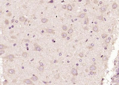
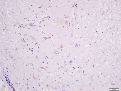
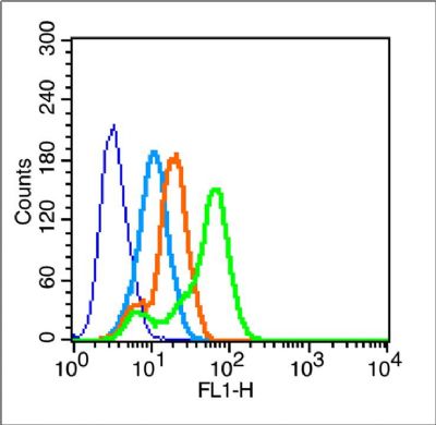
| 英文名称 NMDAR1 中文名称 离子型谷氨酸受体1抗体 别 名 NMDA-NR1; N-Methyl-d-Asprtate receptor 1; GRIN1; NMDA1; NR1; Glutamate [NMDA] receptor subunit zeta 1; Glutamate receptor ionotropic N methyl D aspartate 1; Grin 1; Grin1; N methyl D aspartate receptor channel; N-methyl-D-aspartate receptor; N-methyl-D-aspartate receptor subunit NR1; NMD-R1; NMDA 1; NMDA NR1; NMDA R1; NMDA receptor 1; NMDA1; NMDAR 1; NMDAR; NR 1; NMDZ1_HUMAN. 研究领域 细胞生物 神经生物学 信号转导 细胞凋亡 细胞膜受体 细胞类型标志物 抗体来源 Rabbit 克隆类型 Polyclonal 交叉反应 Human, Mouse, Rat, Chicken, Dog, Pig, Cow, Horse, 产品应用 ELISA=1:500-1000 IHC-P=1:100-500 IHC-F=1:100-500 Flow-Cyt=1μg /test IF=1:100-500 (石蜡切片需做抗原修复) not yet tested in other applications. optimal dilutions/concentrations should be determined by the end user. 分 子 量 103kDa 细胞定位 细胞膜 性 状 Liquid 浓 度 1mg/ml 免 疫 原 KLH conjugated synthetic peptide derived from human NMDAR1:101-200/938 <Extracellular> 亚 型 IgG 纯化方法 affinity purified by Protein A 储 存 液 0.01M TBS(pH7.4) with 1% BSA, 0.03% Proclin300 and 50% Glycerol. 保存条件Shipped at 4℃. Store at -20 °C for one year. Avoid repeated freeze/thaw cycles. PubMedPubMed 产品介绍The protein encoded by this gene is a critical subunit of N-methyl-D-aspartate receptors, members of the glutamate receptor channel superfamily which are heteromeric protein complexes with multiple subunits arranged to form a ligand-gated ion channel. These subunits play a key role in the plasticity of synapses, which is believed to underlie memory and learning. Cell-specific factors are thought to control expression of different isoforms, possibly contributing to the functional diversity of the subunits. Alternatively spliced transcript variants have been described. [provided by RefSeq, Jul 2008] Function: NMDA receptor subtype of glutamate-gated ion channels with high calcium permeability and voltage-dependent sensitivity to magnesium. Mediated by glycine. This protein plays a key role in synaptic plasticity, synaptogenesis, excitotoxicity, memory acquisition and learning. It mediates neuronal functions in glutamate neurotransmission. Is involved in the cell surface targeting of NMDA receptors. Subunit: Forms heteromeric channel of a zeta subunit (GRIN1), a epsilon subunit (GRIN2A, GRIN2B, GRIN2C or GRIN2D) and a third subunit (GRIN3A or GRIN3B); disulfide-linked. Found in a complex with GRIN2A or GRIN2B, GRIN3A or GRIN3B and PPP2CB. Interacts with DLG4 and MPDZ. Interacts with LRFN1 and LRFN2. Interacts with MYZAP. Subcellular Location: Cell membrane; Multi-pass membrane protein. Cell junction, synapse, postsynaptic cell membrane. Cell junction, synapse, postsynaptic cell membrane, postsynaptic density. Note=Enriched in post-synaptic plasma membrane and post-synaptic densities. Post-translational modifications: NMDA is probably regulated by C-terminal phosphorylation of an isoform of NR1 by PKC. Dephosphorylated on Ser-897 probably by protein phosphatase 2A (PPP2CB). Its phosphorylated state is influenced by the formation of the NMDAR-PPP2CB complex and the NMDAR channel activity. DISEASE: Defects in GRIN1 are the cause of mental retardation autosomal dominant type 8 (MRD8) [MIM:614254]. Mental retardation is characterized by significantly below average general intellectual functioning associated with impairments in adaptative behavior and manifested during the developmental period. Similarity: Belongs to the glutamate-gated ion channel (TC 1.A.10.1) family. NR1/GRIN1 subfamily. SWISS: Q05586 Gene ID: 2902 Database links: Entrez Gene: 2902 Human Entrez Gene: 14810 Mouse Entrez Gene: 24408 Rat Omim: 138249 Human SwissProt: Q05586 Human SwissProt: P35438 Mouse SwissProt: P35439 Rat Unigene: 558334 Human Unigene: 278672 Mouse Unigene: 9840 Rat Important Note: This product as supplied is intended for research use only, not for use in human, therapeutic or diagnostic applications. 神经细胞标志物 (NMDAR1)N-甲基-D-天门冬氨酸受体(NMDAR)是兴奋性氨基酸受体亚型,是由NMDAR1与不同的NMDAR2亚基组成的异聚体。 近年实验研究发现,许多NMDAR拮抗药均具有镇痛活性,表明NMDAR在痛觉传递中具有重要作用,这为新型镇痛药的研究开发提供了新的作用靶点。 | 品图片 | Paraformaldehyde-fixed, paraffin embedded (rat brain tissue); Antigen retrieval by boiling in sodium citrate buffer (pH6.0) for 15min; Block endogenous peroxidase by 3% hydrogen peroxide for 20 minutes; Blocking buffer (normal goat serum) at 37°C for 30min; Antibody incubation with (NMDAR1) Polyclonal Antibody, Unconjugated (bs-2030R) at 1:200 overnight at 4°C, followed by operating according to SP Kit(Rabbit) (sp-0023) instructionsand DAB staining. Tissue/cell: rat spinal cord tissue; 4% Paraformaldehyde-fixed and paraffin-embedded; Antigen retrieval: citrate buffer ( 0.01M, pH 6.0 ), Boiling bathing for 15min; Block endogenous peroxidase by 3% Hydrogen peroxide for 30min; Blocking buffer (normal goat serum,C-0005) at 37℃ for 20 min; Incubation: Anti-NMDAR1 Polyclonal Antibody, Unconjugated(bs-2030R) 1:200, overnight at 4°C, followed by conjugation to the secondary antibody(SP-0023) and DAB(C-0010) staining Blank control (blue line): MCF7 (blue).
Primary Antibody (green line): Rabbit Anti-NMDAR1 antibody (bs-2030R)
Dilution: 1μg /10^6 cells;
Isotype Control Antibody (orange line): Rabbit IgG .
Secondary Antibody (white blue line): F(ab’)2 fragment goat anti-rabbit IgG-FITC
Dilution: 1μg /test.
Protocol
The cells were fixed with 2% paraformaldehyde for 10 min at room temperature.Cells stained with Primary Antibody for 30 min at room temperature. The cells were then incubated in 1 X PBS/2%BSA/10% goat serum to block non-specific protein-protein interactions followed by the antibody for 15 min at room temperature. The secondary antibody used for 40 min at room temperature. Acquisition of 20,000 events was performed. | |